

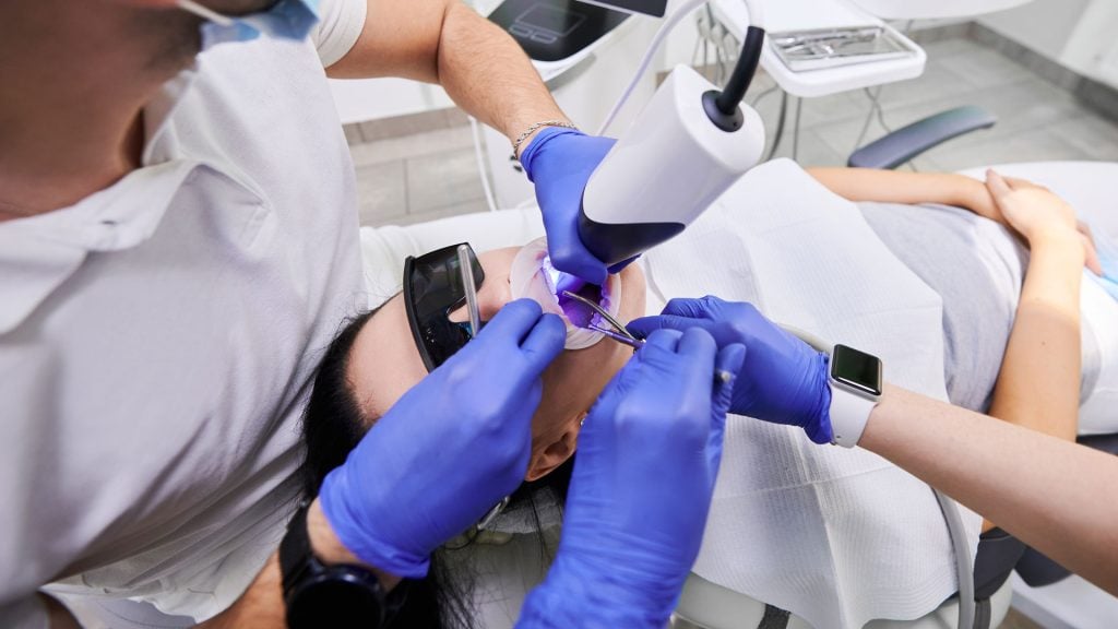
Intra Oral Camera
Dental Technology
Intra Oral Camera is a small video camera that allows us to photograph any areas of concern in your mouth – both periodontal and restorative. By seeing and understanding what the dentist sees, you are better able to understand and therefore make informed decisions about your dental health treatment.
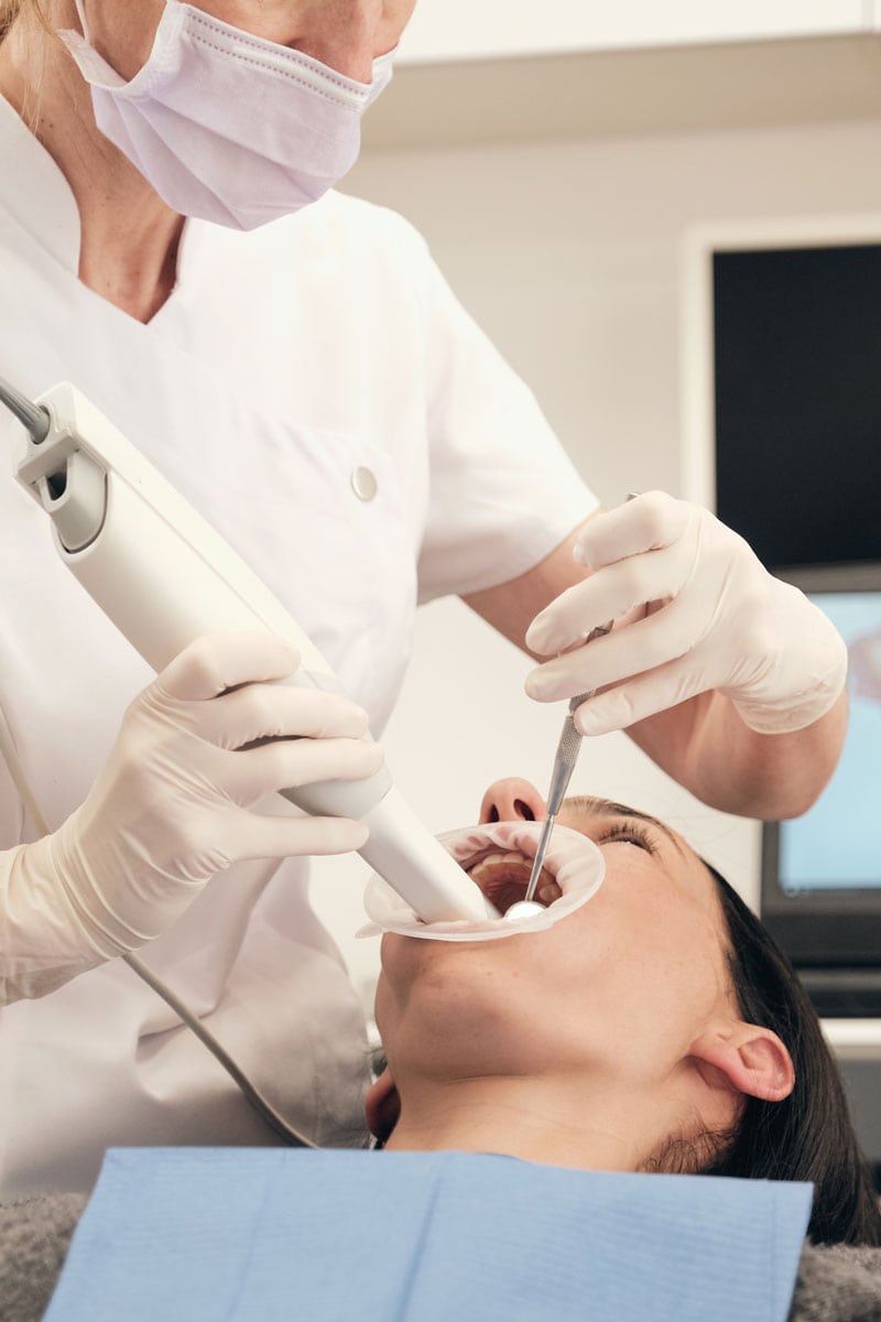
Intra Oral Camera at Arden Park Dental
An intraoral camera is a small, handheld device used by dentists to capture detailed, high-resolution images of the inside of your mouth. This camera is a valuable diagnostic and educational tool in modern dentistry, providing both the dentist and the patient with a clearer, more accurate view of the oral cavity.
An intraoral camera is a powerful and efficient tool in modern dentistry that helps enhance diagnosis, improve patient education, and streamline treatment planning.
By allowing both the dentist and the patient to visually inspect and understand oral health, the camera can lead to better communication and outcomes. It also helps detect problems early, ensuring that treatments are more effective and minimally invasive.
How Digital X-rays Work
Small, Handheld Device
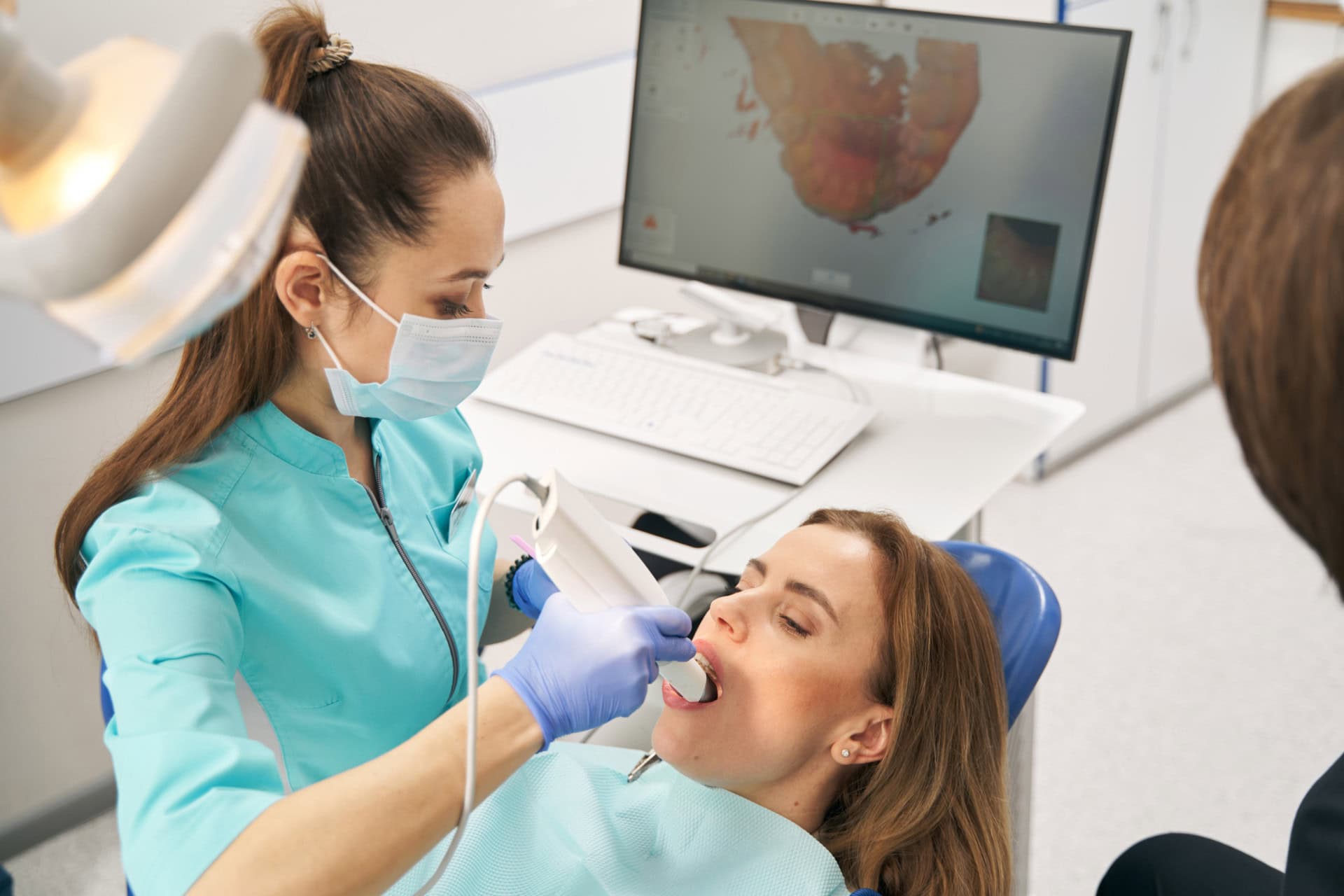
Digital Imaging
Light Source
Real-Time Visualization
Enhanced Diagnosis and Treatment Planning
- The images produced by an intraoral camera can help the dentist identify issues early that might be missed with the naked eye, such as small cavities, gum disease, cracks in teeth, tooth decay, or broken fillings.
- It allows for more accurate diagnoses by providing a detailed view of hard-to-reach areas, such as the back of molars or the margins of fillings and crowns.
Visual Education for Patients
- One of the biggest advantages of intraoral cameras is their ability to educate patients. By seeing clear images of their teeth and gums on a monitor, patients can better understand their dental health, what issues need to be addressed, and why certain treatments are necessary.
- For example, if a patient has a cavity or gum disease, the dentist can show them the exact location of the problem, which can make the patient feel more involved in the treatment process and help them understand the importance of care recommendations.
Improved Communication
- Intraoral cameras make it easier for the dentist to communicate with patients. Instead of relying solely on verbal explanations or X-ray images, which can be hard to interpret, the dentist can show real-time pictures of problem areas in the mouth, making the conversation clearer and more impactful.
- Many dental insurance companies now accept intraoral images as part of treatment documentation, which can be important for approval of claims.
Documentation and Monitoring
- Images captured by the camera can be stored digitally, allowing the dentist to track changes over time. For example, the dentist can compare images taken at different appointments to monitor the progression of tooth decay, the effectiveness of treatments, or the healing of gum tissue.
- This provides both the dentist and the patient with a clear record of oral health that can be referenced as needed.
Minimally Invasive
- The intraoral camera is non-invasive and painless. Unlike X-rays, which require radiation, the camera only needs to be positioned in the mouth to capture images and does not involve any discomfort or risk to the patient.
Efficiency
- Since the images are captured in real-time and displayed instantly, intraoral cameras help make dental visits faster and more efficient. There’s no need to wait for X-rays to be developed or processed, which can speed up diagnosis and treatment planning.
Early Detection of Problems
- With the ability to magnify and illuminate areas in the mouth, an intraoral camera can detect early signs of cavities, gum inflammation, oral cancer, and other oral health issues before they become more serious or painful. Early intervention can often result in less invasive and more cost-effective treatments.
- Detecting Cavities and Tooth Decay:
- Close Examination: Intraoral cameras allow the dentist to closely examine teeth for early decay that may not be visible to the naked eye. The high-resolution images can highlight small areas of damage that can be treated before they become larger cavities.
- Monitoring Gum Health:
- Gum Inspection: The camera can be used to visually inspect the gums and detect signs of gum disease, such as inflammation, infection, or receding gums. It can also show the condition of the tissue around dental implants or around fillings and crowns.
- Examine Fillings, Crowns, and Other Restorations:
- Checking Existing Dental Work: Intraoral cameras are useful for assessing the condition of existing dental work, such as fillings, crowns, bridges, and implants. They help ensure that restorations are intact and functioning well, and can reveal any cracks or wear on the materials.
- Detecting Cracks and Fractures:
- High-Resolution Imaging: Small cracks or fractures in teeth can sometimes be hard to detect, but the high-resolution imaging from an intraoral camera can capture them clearly. This is important for avoiding future problems that could lead to more extensive damage or the need for root canal therapy.
- Patient Education and Treatment Planning:
- Visual Understanding: Intraoral cameras allow patients to see exactly what the dentist sees. This helps patients understand their diagnosis better and make informed decisions about their treatment options.
- Motivation for Treatment: For example, when a patient sees a visible cavity on a tooth or receding gums, they may be more likely to follow the dentist’s recommendations for restorative or preventive treatments.
- Oral Cancer Screening:
- Identifying Abnormalities: The camera can be used to identify signs of oral cancer by closely examining the tissues of the mouth, tongue, gums, and cheeks. It can help detect abnormal tissue changes, lesions, or suspicious spots that could require further investigation.
- Tracking and Documenting Treatment Progress:
- Visual Progress Reports: For patients undergoing long-term treatments (like braces or gum therapy), intraoral cameras can help document treatment progress by capturing images before, during, and after treatment.
Limitations of the Intraoral Camera
Limited Field of View
While intraoral cameras are excellent for close-up, detailed images of the teeth and gums, their field of view is limited to the small area in front of the camera. They can’t provide a comprehensive, panoramic view of the mouth like X-rays or 3D imaging.
Not a Substitute for X-rays
Intraoral cameras are a great diagnostic tool, but they can’t replace X-rays or other imaging techniques that provide a broader view of underlying structures, such as the bone, roots of the teeth, and areas that are hidden beneath the gum line.
Skill and Experience Required
The effectiveness of the intraoral camera depends on the skill and experience of the dental professional using it. While the camera provides high-resolution images, it’s up to the dentist to interpret them correctly and use the information to guide treatment.
Patient Sensitivity
Some patients may feel uncomfortable or experience a gag reflex when the camera is placed in the back of the mouth, although the camera is usually small and easy to maneuver.